

Palabras clave
Cómo citar
Resumen
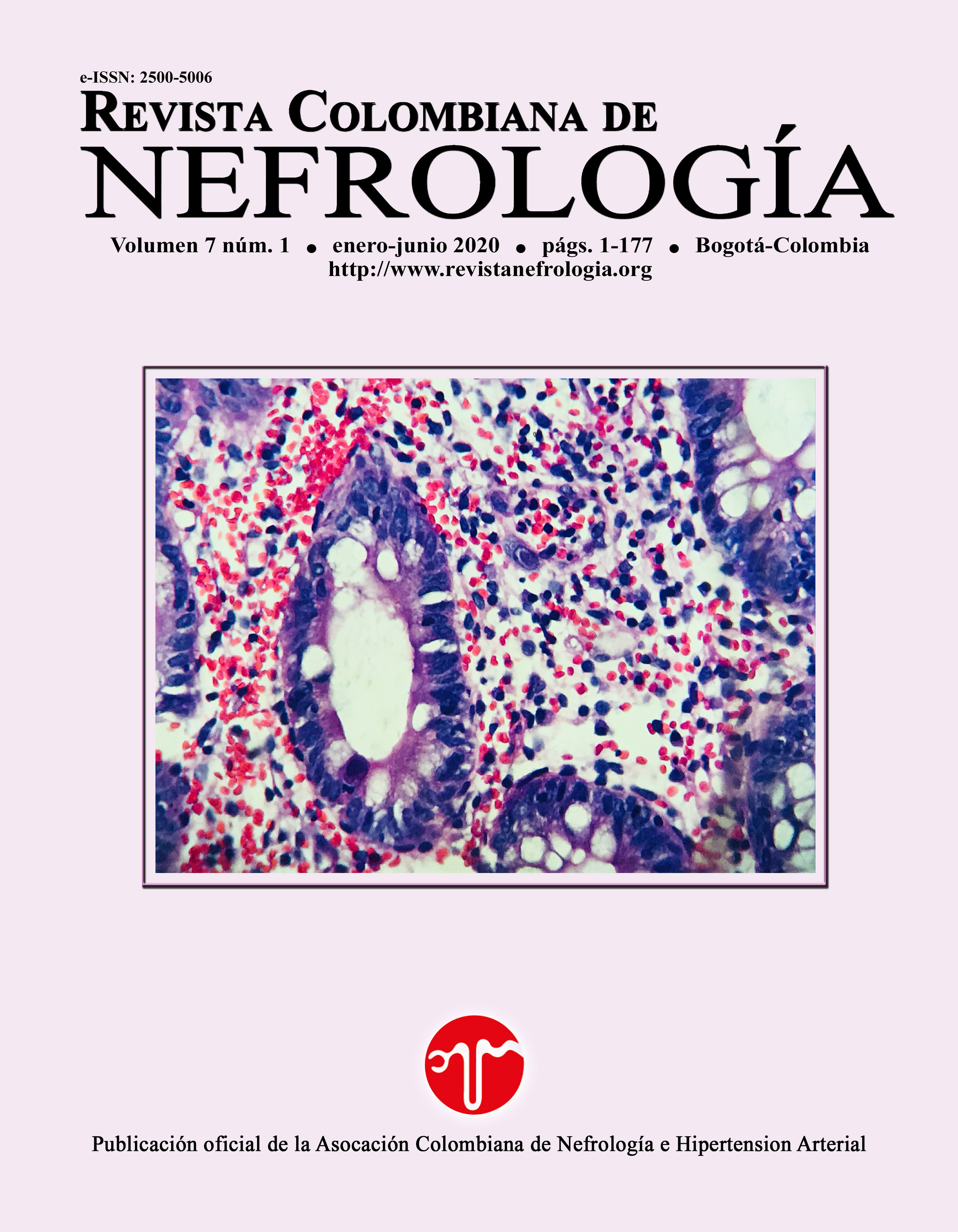
En pacientes con enfermedad renal se ha reportado la presencia de células renales reactivas, cuyas alteraciones morfológicas severas dificultan su clasificación e interpretación. El conocimiento de las características morfológicas y los patrones de sedimentos en donde se presentan pueden ser de ayuda para su manejo en los departamentos médicos correspondientes. Aquí, nosotros reportamos la presencia de células agrupadas en acinos, con abundante citoplasma, cariomegalia, contornos nucleares irregulares y nucléolos prominentes, acompañados de cilindruria y cuerpos ovales grasos en el sedimento urinario de dos pacientes con diabetes mellitus, las cuales fueron sugestivas de células renales reactivas.
Citas
Ross MH, Pawlina W. Histología. Texto y Atlas. 7 ed. Barcelona, España: Wolters Kluwer; 2015.
Koss LG, Hoda RS. Koss’s cytology of the urinary tract with histopathologic correlations. Springer; 2012.
Khandelwal P, Abraham SN, Apodaca G. Cell biology and physiology of the uroepithelium. Am J Physiol Renal Physiol. 2009;297(6):1477-501. Available from: https://doi.org/10.1152/ajprenal.00327.2009
JCCLS. Urinary Sediment Examination. Japanese J Med Technol. 2017;66:51-85. Available from: https://doi.org/10.14932/jamt.17J1-2e
Fogazzi GB. The urinary sediment. An integrated view. 3 ed. Italia: Elsevier; 2010.
Nguyen GK, Smith R. Repair renal tubular cells: A potential false-positive diagnosis in urine cytology. Diagn Cytopathol. 2004;31(5):342-6. Available from: https://doi.org/10.1002/dc.20139
Ohsaki H, Hirakawa E, Kushida Y, Yokoshita S, Nakamura M, Kiyomoto H, et al. Can cytological features differentiate reactive renal tubular cells from low-grade urothelial carcinoma cells? Cytopathology. 2010;21(5):326-33. Available from: https://doi.org/10.1159/000325510
Cruz Abascal RE, Fuentes Flebes O, Gutiérrez Simón O, Garay Padrón R, Águila Moya O. Nefropatía diabética en pacientes diabéticos tipo 2. Rev Cuba med. 2011;50(1):29-39.
González Álvarez MT, Mallafré Anduig J. Nefrología. Conceptos básicos en atención primaria. 1a ed. Barcelona, España: MARGE MEDICA BOOKS; 2009.
Ohsaki H, Haba R, Matsunaga T, Nakamura M, Kiyomoto H, Hirakawa E. Cytomorphologic and inmmunocytochemical characteristics of reactive renal tubular cells in renal glomerular disease. Acta Cytol. 2008;52(3):297-301. Available from: https://doi.org/10.1159/000325510
Caleffi A, Lippi G. Cylindruria. Clin Chem Lab Med. 2015;53(s2):1471-7. Available from: https://doi.org/10.1515/cclm-2015-0480
Muto S, Sugiura S, Nakajima A, Horiuchi A, Inoue M, Saito K, et al. Isomorphic red blood cells using automated urine flow cytometry is a reliable method in diagnosis of bladder cancer. Int J Clin Oncol. 2014;19(5):928-34. Available from: https://doi.org/10.1007/ s10147-013-0623-9
Fogazzi GB, Edefonti A, Garigali G, Giani M, Zolin A, Raimondi S, et al. Urine erythrocyte morphology in patients with microscopic haematuria caused by a glomerulopathy. Pediatr Nephrol. 2008;23(7):1093-100. Available from: https://doi.org/10.1007/s00467-008-0777-2
Quinn JR, Zimmerman HJ. Significance of Oval Fat Bodies in Urinary Sediment. Am J Clin Pathol. 1954;24(7):787-95. Available from: https://doi.org/10.1093/ajcp/24.7.787
Strojan M, Srebotnik I, Gutnik H. Use of Vimentin Immunocytochemical Staining for Evaluation of Atypical Cells in Voided Urine Samples. Diagn Cytopathol. 2016;45(2):85-90. Available from: https://doi.org/10.100/dc.23645134
- Los autores/as conservarán sus derechos de autor y garantizarán a la revista el derecho de primera publicación de su obra, el cuál estará simultáneamente sujeto a la Licencia de Creative Commons Reconocimiento- NoComercial- SinObraDerivada 4.0 Internacional. que permite a terceros compartir la obra siempre que se indique su autor y su primera publicación esta revista.